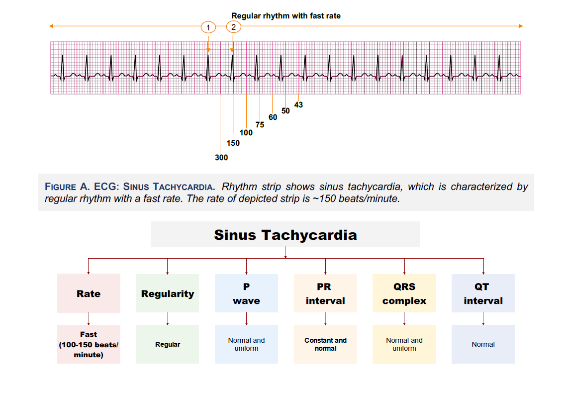
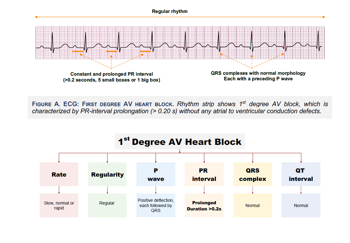
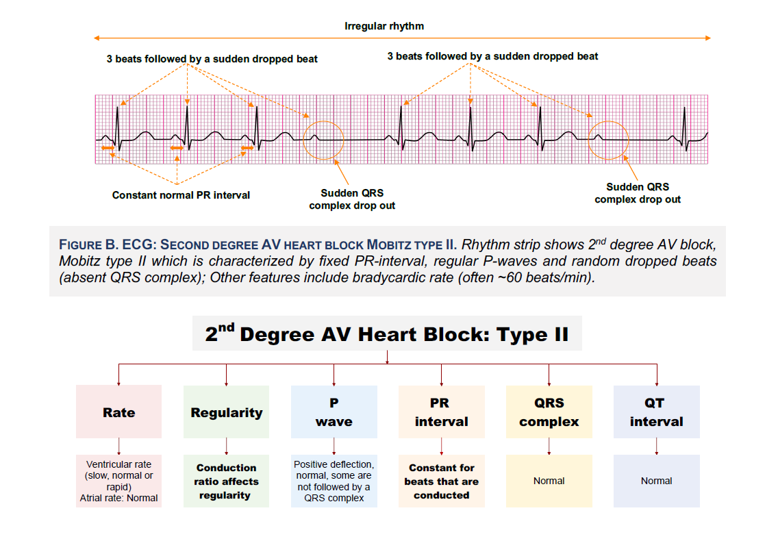
Systematic Approach to EKG and Interpretation for COMLEX Level 2 CE

Systematic Approach to EKG and Interpretation for COMLEX Level 2 CE

The material contains detailed information for anatomy and physiology, using a heart monitor, as well as identifying and interpreting EKG tracings, including:

  • Recognizing sinus rhythms
  • Cardiac & pulmonary syndromes
  • Sinus node, atrial, junctional, and ventricular arrhythmias
  • AV conduction abnormalities
  • Pacemakers and ICD
  • Drug side effects
  • Bundle Branch Blocks
  • 3-lead, 5-lead, and 12-lead EKG tracings
  • In-depth diagrams for electrode placement
  • Electrolyte imbalances & metabolic factors
  • Procedures for commonly performed tests

Study abnormal EKG tracings for COMLEX Level 2 CE

Study abnormal EKG tracings for COMLEX Level 2 CE

The EKG review discusses a systematic approach to rhythm interpretation to help diagnose, monitor, and determine the severity of conditions such as:

  • -Congenital heart defects
  • -Coronary artery disease
  • -Heart attack
  • -Heart failure
  • -Heart murmurs
  • -Cardiomyopathy
  • -Pericarditis
  • -Cardiac arrhythmias

Study EKG patterns and cardiac structural abnormalities for COMLEX Level 2 CE

Study EKG patterns and cardiac structural abnormalities for COMLEX Level 2 CE

Improve your ability to accurately interpret EKG tracings for irregularities and abnormalities which is a core competency for COMLEX Level 2 CE.

Identify conditions caused by malfunctions in the heart’s electrical system from medication and drug use such as amphetamines, OTC cold and allergy meds, beta blockers, and cocaine.

Learn how to recognize structural abnormalities that give clues to potential heart problems or defects such as enlargement of the chambers or walls

Learn EKG Conduction Abnormalities for COMLEX Level 2 CE

Learn the art of EKG interpretation through an organized approach to make the process more simple for COMLEX Level 2 CE

Classification of supraventricular tachycardia

Visually rich Qbank graphics

More than 3,000 highly detailed graphics, tables, diagrams, flow charts and illustrations.

The UMock methodology helps organize the underlying basic concepts with a high quality presentation of visual aids.

The use of visual aids from UMock makes it easier to understand complex concepts, improve retention, and reduce study time.

COMLEX Level 2 Plans

Course Content

Expand All

● Cardiac Muscle

● Cardiac Chambers

● Coronary Artery Anatomy

● Cardiac Conduction System

● Cardiac Action Potentials

● Cardiac Innervation

● Cardiac Cycle

● Electrocardiogram

● ECG Electrodes

● Limb Leads

● Precordial Leads

● Contiguous Leads

● ECG Paper

● ECG Recording

● ECG Waves

● ECG Segments and Intervals

● Heart Rate Determination

● Cardiac Axis Determination

● Electrode Misplacement

● P Wave

● PR Interval

● QRS Complex

● R Wave Progression

● ST Segment

● T Wave

● QT Interval

● U Wave

● Atrial Enlargement

● Ventricular Enlargement

● Classification of Arrhythmias

● Tachyarrhythmias

● AV Nodal Reentry Tachycardia (AVNRT)

● Classification of AVNRT

● Atrioventricular Reentry Tachycardias (AVRT)

● Triggered Activity

● Wide QRS Tachyarrhythmias

● VT versus SVT

● Vereckei Algorithm

● Brugada Algorithm

● Adenosine

● Adenosine Effect on Arrhythmias

● Sinus Tachycardia

● Sinus Bradycardia

● Sinus Dysrhythmias

● Sinus Arrest and Pause

● Sick Sinus Syndrome

● Atrial Arrhythmias

● Wandering Atrial Pacemaker

● Premature Atrial Complexes

● First Degree Heart Block

● Second Degree Heart Block

● Third Degree Heart Block

● Atrial Flutter

● Atrial Fibrillation

● Atrial Tachycardia

● Multifocal Atrial Tachycardia

● Premature Junctional Beat

● Early Repolarization

● Wolf-Parkinson-White Syndrome

● Junctional Tachycardia

● Benign Early Repolarization

● Ventricular Arrhythmias

● Premature Ventricular Complexes

● Couplets and Bigeminy

● Trigeminy

● Idioventricular Rhythm

● Ventricular Tachycardia

● Torsade de Pointes

● Ventricular Fibrillation

● Asystole

● Hyperkalemia

● Hypokalemia

● Hypercalcemia

● Hypocalcemia

● Hyperthyroidism

● Hypothyroidism

● Digitalis Effect

● ß-Blocker Toxicity

● Sotalol Toxicity

● Calcium Channel Blockers Toxicity

● Quetiapine Toxicity

● Electrical Alternans

● Pericarditis

● Pulmonary Embolism

● Angina

● Acute Coronary Syndrome

● Myocardial Infarction

● STEMI

● Sudden Cardiac Death

● Hypertrophic Cardiomyopathy

● Cardiac Muscle

● Cardiac Action Potential

● WolfParkinson-White Syndrome

● Hypothermia

● Wellen’s Syndrome

● Myocardial Infarction

● ACS Syndrome

● STEMI

● Pulmonary Embolism

● Brainstem Herniation

● De Winter’s ECG Sign

● Sotalol Toxicity

● Propranolol Toxicity

● Tricyclic Antidepressant Toxicity

EKG Book Content

COMLEX Level 2 EKG Interpretation

Prepare for your exam with UMock EKG Review. Each chapter includes a
complete review of topics with a Q&A at the end of the book.

Sinus Rhythms
Learn how to identify features and qualifying criteria of sinus rhythms including normal, sinus bradycardia, sinus tachycardia, sinus dysrhythmia, sinus arrest, and sinus exit block.
Atrial Rhythms
Recognize complexes and rhythms for premature atrial complexes, wandering atrial pacemaker rhythm, multifocal atrial tachycardia, atrial flutter, and atrial fibrillation.
Junctional Rhythms
Identify features and criteria for premature junctional complexes, junctional escape beats, accelerated junctional rhythm, junctional tachycardia, and supraventricular tachycardia.
Heart Block Rhythms
Review the features and criteria for first degree heart block, second degree heart block type I, second degree heart block type II, and third degree/complete heart block.
Ventricular Rhythms
Identify features of ventricular rhythms including premature ventricular complexes, agonal rhythm, idioventricular and accelerated idioventricular rhythm, ventricular tachycardia, ventricular fibrillation, and asystole.
Pacemaker Rhythms
Learn how to recognize features of pacemaker rhythms including atrial and ventricular pacemaker rhythm, atrioventricular pacemaker rhythm, and failure to capture.
12-Lead ECG Axis
Review of which major coronary artery supports each area of the heart and learn the fundamentals of electrical axis determination and the presence of cardiac axis deviation.
Performing 12-Lead
Learn 12-lead EKG tracings which presents the electrical activity of the heart on two planes. Leads I, II, III, aVR, aVL, and aVF give a flat view of the heart from surrounding edges of a frontal plane.
Bundle Branch Block
Recognize the features for the presence of a complete left or right bundle branch block.
Left Ventricular Hypertrophy
Learn how to recall and apply the process for measurement to determine the presence of left ventricular hypertrophy.
Injury, Ischemia & Infarction
Review of the fundamentals and differences of indicative and reciprocal views, and recognize EKG changes caused by STEMI, ischemia, injury, and infarction.
EKG Terminology
Learn basic EKG terminology including PR interval, QRS complex, QT interval and ST interval.

" The EKG review from UMock provides a comprehensive overview and complete understanding interpreting tracings for every possible heart condition. The content covers everything you’ll see on Level 2 CE related to analyzing and interpreting tracings. "

Natalie

University of California, Los Angeles

0